
The best time to do a 3D ultrasound is generally between 26 to 30 weeks of pregnancy. This timeframe is recommended because, by this stage, the baby has developed sufficient body fat, making their facial features and body more distinguishable and detailed in the images.
Before 26 weeks, the baby might appear too lean, making it difficult to see clear features. After 30 weeks, it becomes more challenging to get clear images due to the limited space in the womb as the baby grows larger and has less room to move around. This period offers a good balance, providing clear, detailed images while still having enough space around the baby to capture different angles and positions.
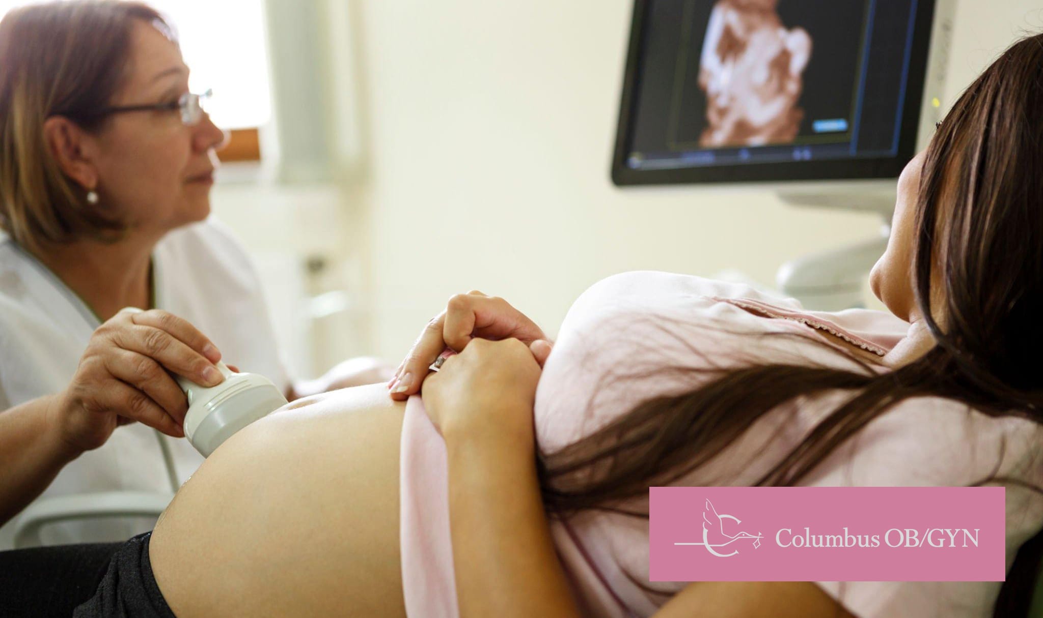
An ultrasound is a machine emitting sound waves, which are not to be confused with x-rays. It uses these sound waves to create images of your baby within the womb. A small device called a transducer is placed on the mother’s abdomen, emitting high-frequency sound waves that bounce off the baby’s surfaces, including internal organs and bones. These echoes are captured and translated onto a computer screen as visual images.
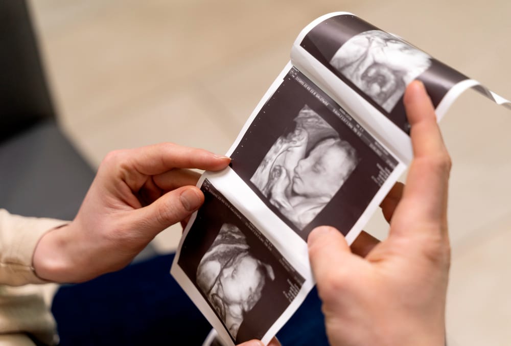
For expectant parents looking for more clarity and connection during pregnancy, advanced ultrasounds, like 3D ultrasounds go a step further by taking thousands of images at once, compiling them into a three-dimensional image that offers a much clearer view of the baby’s features compared to the flat, two-dimensional images produced by standard ultrasounds.
Getting an ultrasound is an exciting part of the pregnancy process, but not every ultrasound is appropriate for every step of the baby’s development. There are so many reasons why your doctor might recommend an ultrasound; here’s the breakdown of what pregnant women can expect for a look at what’s occurring inside the womb:
Dating and Viability Ultrasound: Done to confirm the pregnancy, estimate the due date, and check the number of embryos. It can also assess the pregnancy’s health and viability.
Nuchal Translucency Screening: Part of the screening for chromosomal abnormalities like Down syndrome. This measures the fluid at the back of the baby’s neck and monitors the amniotic fluid surrounding the entire baby.
Anatomy Scan (Level 2 Ultrasound): Conducted by a Hilliard Gynecologist one to two weeks before or after the 20-week mark to examine the fetus’s anatomy in detail, including the brain, heart, bones, and organs, to check for any developmental abnormalities, such as birth defects. This is also a common time when expecting parents can learn the baby’s sex if they choose to.
Follow-up Ultrasounds: May be required for any specific issues identified or for high-risk pregnancies to monitor the baby’s development closely. These ultrasounds are particularly important for expectant parents or mothers worried about potential complications.
Several factors influence whether your 3D ultrasound will result in detailed images:
Aside from keepsake images, 3D ultrasounds have specific medical purposes. Providers may use 3D technology to:
The detailed nature of these scans makes them useful when routine ultrasounds raise concerns.
The optimal window to get a 3D ultrasound is generally in the third trimester, between 26 to 30 weeks of pregnancy. During this period, your baby has developed enough fat under their skin to show distinct facial features, yet still has enough space to move around, which is necessary for capturing different angles and postures.
Before 26 weeks pregnant, your baby might look a bit lean in the ultrasound images, as they haven’t developed much body fat yet. After 30 weeks, the increasing crowding in the womb makes it more challenging to get clear images, as the baby has less room to move into a position that would be conducive to get a 3D ultrasound.
It’s also important to follow the advice of your gynecologist in Lewis Center regarding the number and timing of any ultrasound. While 3D ultrasounds are generally safe, they should be performed by qualified professionals in a clinical setting to ensure both mother and baby’s safety.
In addition to scheduling your 3D ultrasound, it’s essential to prioritize prenatal care during your pregnancy. Regular visits to your OB/GYN will ensure that both you and your baby are healthy and that any concerns can be addressed early.
Columbus OB/GYN proudly provides safe and advanced 3D ultrasound services to families across Columbus, Grandview, Dublin, and Hilliard—helping you experience those special moments with clarity and peace of mind.
No, there is no evidence to suggest that the sound waves used in ultrasounds to obtain images of the unborn child, including 3D ultrasounds, harm the baby. Ultrasounds are considered a safe and non-invasive method for monitoring the health and development of the fetus.
The best time to see the baby’s facial features clearly on a 3D ultrasound is between 26 to 30 weeks of pregnancy. This is when there is enough facial fat on the baby’s face to show features like the nose, lips, and eyes more clearly.